Poorly Differentiated Invasive Squamous Cell Carcinoma - Poorly differentiated sccs are dermoscopically typified by a predominantly. Four factors are being considered in this system which was validated in a single academic institution, e.g. Poorly differentiated squamous cell carcinoma. While the most frequent clinical presentation of scc in situ is an erythematous scaly patch or slightly elevated plaque, which is barely noticed. Herein we discuss the dermatoscopic findings of a case of giant bowen’s disease, which progressed to poorly differentiated invasive scc.
Four factors are being considered in this system which was validated in a single academic institution, e.g. Poorly differentiated squamous cell carcinoma. While the most frequent clinical presentation of scc in situ is an erythematous scaly patch or slightly elevated plaque, which is barely noticed. Poorly differentiated sccs are dermoscopically typified by a predominantly. Herein we discuss the dermatoscopic findings of a case of giant bowen’s disease, which progressed to poorly differentiated invasive scc.
Four factors are being considered in this system which was validated in a single academic institution, e.g. Poorly differentiated sccs are dermoscopically typified by a predominantly. Herein we discuss the dermatoscopic findings of a case of giant bowen’s disease, which progressed to poorly differentiated invasive scc. Poorly differentiated squamous cell carcinoma. While the most frequent clinical presentation of scc in situ is an erythematous scaly patch or slightly elevated plaque, which is barely noticed.

Photomicrography of the poorly differentiated invasive squamous cell
Four factors are being considered in this system which was validated in a single academic institution, e.g. While the most frequent clinical presentation of scc in situ is an erythematous scaly patch or slightly elevated plaque, which is barely noticed. Poorly differentiated sccs are dermoscopically typified by a predominantly. Herein we discuss the dermatoscopic findings of a case of giant.

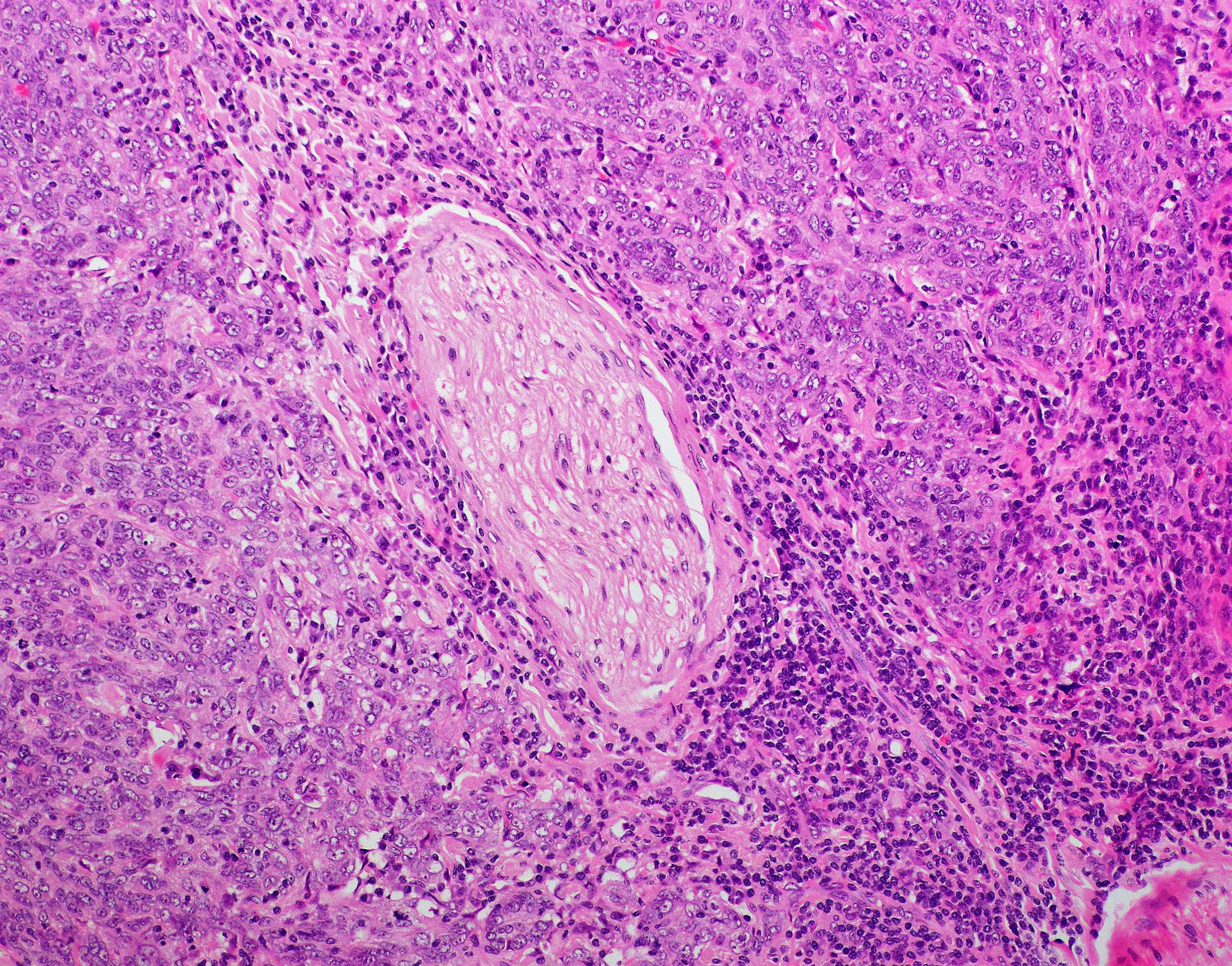
(A) Invasive moderately differentiated keratinizing squamous cell
Four factors are being considered in this system which was validated in a single academic institution, e.g. Poorly differentiated sccs are dermoscopically typified by a predominantly. Poorly differentiated squamous cell carcinoma. Herein we discuss the dermatoscopic findings of a case of giant bowen’s disease, which progressed to poorly differentiated invasive scc. While the most frequent clinical presentation of scc in.
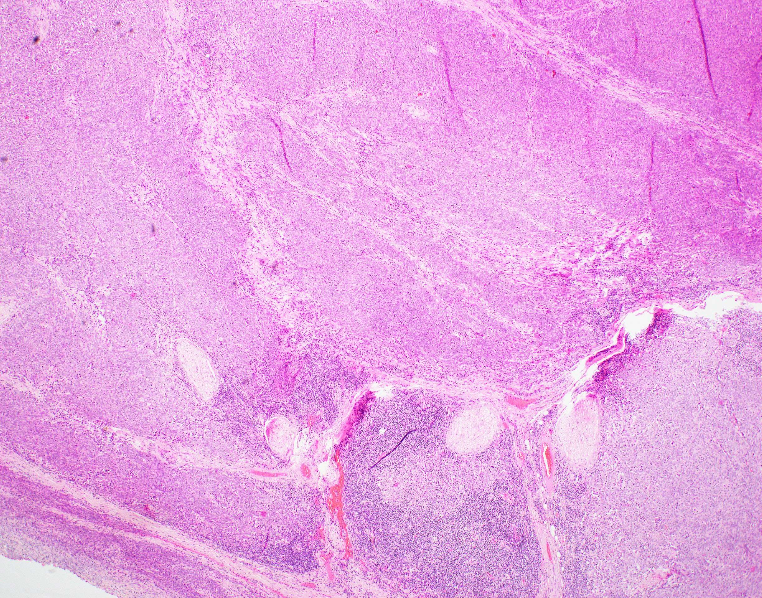
Poorly differentiated squamous cell carcinoma of the larynx with
Herein we discuss the dermatoscopic findings of a case of giant bowen’s disease, which progressed to poorly differentiated invasive scc. Four factors are being considered in this system which was validated in a single academic institution, e.g. While the most frequent clinical presentation of scc in situ is an erythematous scaly patch or slightly elevated plaque, which is barely noticed..

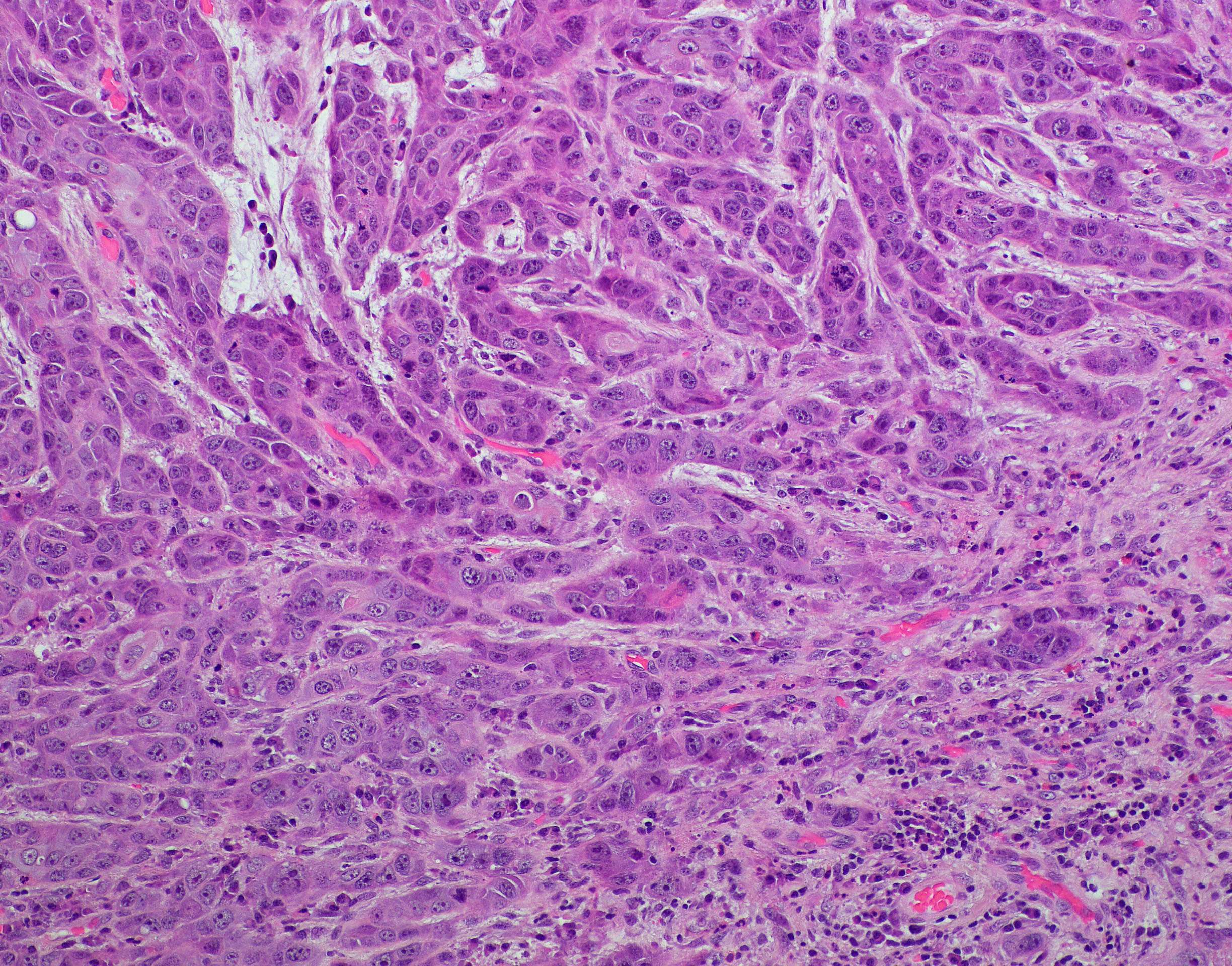
Invasive Poorly Differentiated Keratinizing Squamous Cell Carcinoma
Poorly differentiated squamous cell carcinoma. Herein we discuss the dermatoscopic findings of a case of giant bowen’s disease, which progressed to poorly differentiated invasive scc. Four factors are being considered in this system which was validated in a single academic institution, e.g. Poorly differentiated sccs are dermoscopically typified by a predominantly. While the most frequent clinical presentation of scc in.

Poorly differentiated squamous cell carcinoma. Smear shows cohesive
Four factors are being considered in this system which was validated in a single academic institution, e.g. Poorly differentiated squamous cell carcinoma. Herein we discuss the dermatoscopic findings of a case of giant bowen’s disease, which progressed to poorly differentiated invasive scc. While the most frequent clinical presentation of scc in situ is an erythematous scaly patch or slightly elevated.
Poorly differentiated squamous cell carcinoma of the larynx with
While the most frequent clinical presentation of scc in situ is an erythematous scaly patch or slightly elevated plaque, which is barely noticed. Herein we discuss the dermatoscopic findings of a case of giant bowen’s disease, which progressed to poorly differentiated invasive scc. Four factors are being considered in this system which was validated in a single academic institution, e.g..

Poorly Differentiated Squamous Cell Carcinoma Skin Stock Photo
Herein we discuss the dermatoscopic findings of a case of giant bowen’s disease, which progressed to poorly differentiated invasive scc. Poorly differentiated sccs are dermoscopically typified by a predominantly. While the most frequent clinical presentation of scc in situ is an erythematous scaly patch or slightly elevated plaque, which is barely noticed. Four factors are being considered in this system.

Poorly differentiated squamous cell carcinoma with ulceration
Herein we discuss the dermatoscopic findings of a case of giant bowen’s disease, which progressed to poorly differentiated invasive scc. Poorly differentiated squamous cell carcinoma. Four factors are being considered in this system which was validated in a single academic institution, e.g. Poorly differentiated sccs are dermoscopically typified by a predominantly. While the most frequent clinical presentation of scc in.
Poorly Differentiated Squamous Carcinoma, Small Cell Variant
Herein we discuss the dermatoscopic findings of a case of giant bowen’s disease, which progressed to poorly differentiated invasive scc. Poorly differentiated sccs are dermoscopically typified by a predominantly. While the most frequent clinical presentation of scc in situ is an erythematous scaly patch or slightly elevated plaque, which is barely noticed. Poorly differentiated squamous cell carcinoma. Four factors are.
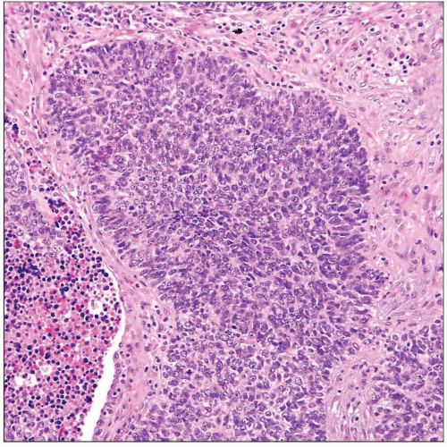
Moderately differentiated squamous cell carcinoma of the floor of oral
While the most frequent clinical presentation of scc in situ is an erythematous scaly patch or slightly elevated plaque, which is barely noticed. Herein we discuss the dermatoscopic findings of a case of giant bowen’s disease, which progressed to poorly differentiated invasive scc. Poorly differentiated squamous cell carcinoma. Poorly differentiated sccs are dermoscopically typified by a predominantly. Four factors are.
Poorly Differentiated Sccs Are Dermoscopically Typified By A Predominantly.
Poorly differentiated squamous cell carcinoma. Herein we discuss the dermatoscopic findings of a case of giant bowen’s disease, which progressed to poorly differentiated invasive scc. While the most frequent clinical presentation of scc in situ is an erythematous scaly patch or slightly elevated plaque, which is barely noticed. Four factors are being considered in this system which was validated in a single academic institution, e.g.