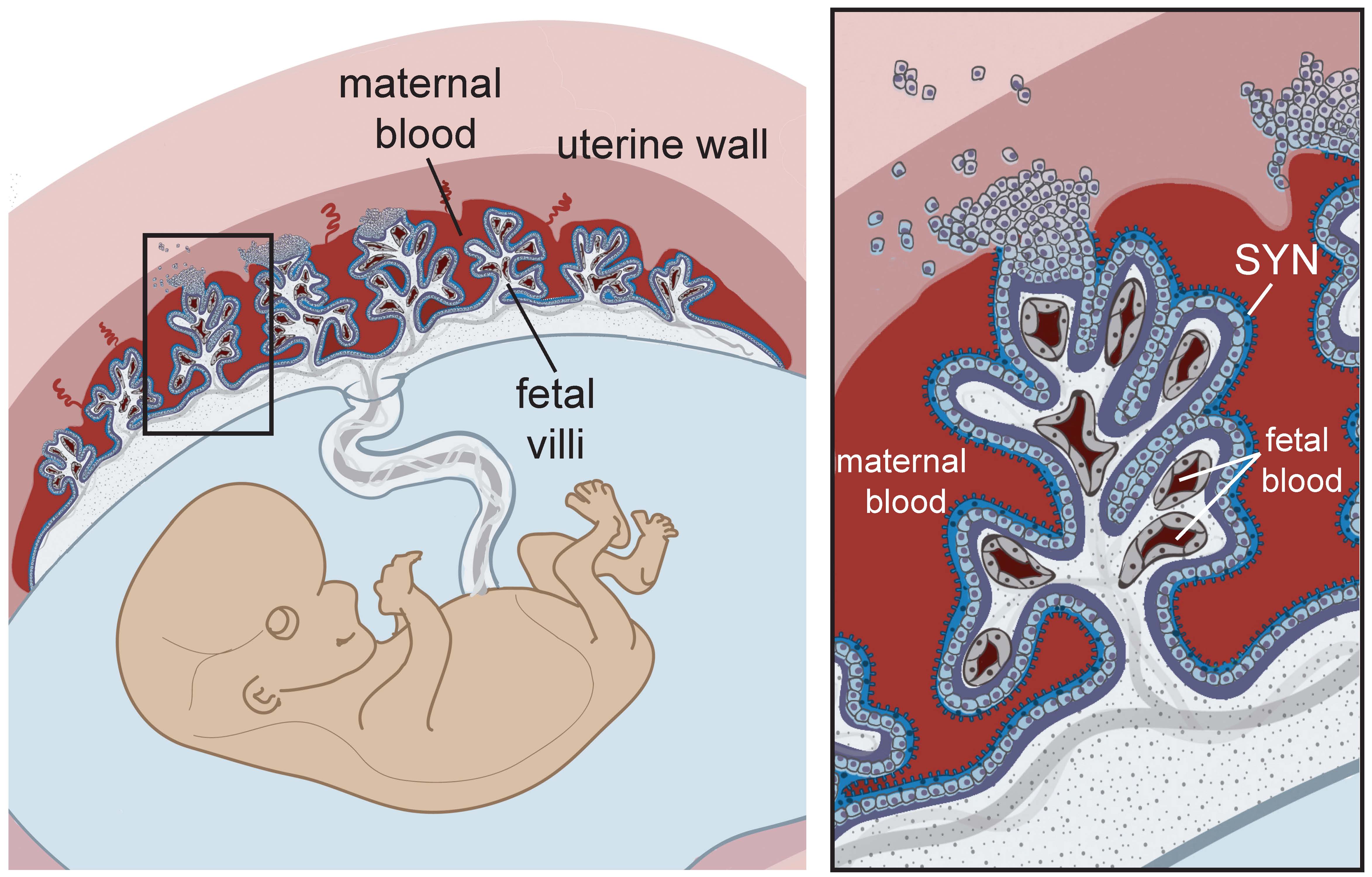
Placenta And Amniotic Sac Diagram - In placenta increta, villi penetrate deeper into the myometrium. In placenta percreta, villi reach the uterine serosa and/or invade. The placental amnion [ figure 1a (1)] covers the placenta, and the reflected amnion [ figure 1a (2)] lines the uterine wall, thereby forming the. Here’s a detailed description of the umbilical cord, placenta, amniotic sac, and amniotic fluid, including their functions in the. The amnion, umbilical cord, and fetal vessels are visible. The placenta is a connection between foetal membrane and the inner uterine wall. Download scientific diagram | anatomy of human placenta. Thus, placenta is partly maternal and partly. The fetus developing within the amniotic sac is surrounded and encased by trophoblast cells in the placenta and chorion membrane (red).
The fetus developing within the amniotic sac is surrounded and encased by trophoblast cells in the placenta and chorion membrane (red). In placenta increta, villi penetrate deeper into the myometrium. The amnion, umbilical cord, and fetal vessels are visible. Download scientific diagram | anatomy of human placenta. Thus, placenta is partly maternal and partly. Here’s a detailed description of the umbilical cord, placenta, amniotic sac, and amniotic fluid, including their functions in the. The placenta is a connection between foetal membrane and the inner uterine wall. In placenta percreta, villi reach the uterine serosa and/or invade. The placental amnion [ figure 1a (1)] covers the placenta, and the reflected amnion [ figure 1a (2)] lines the uterine wall, thereby forming the.
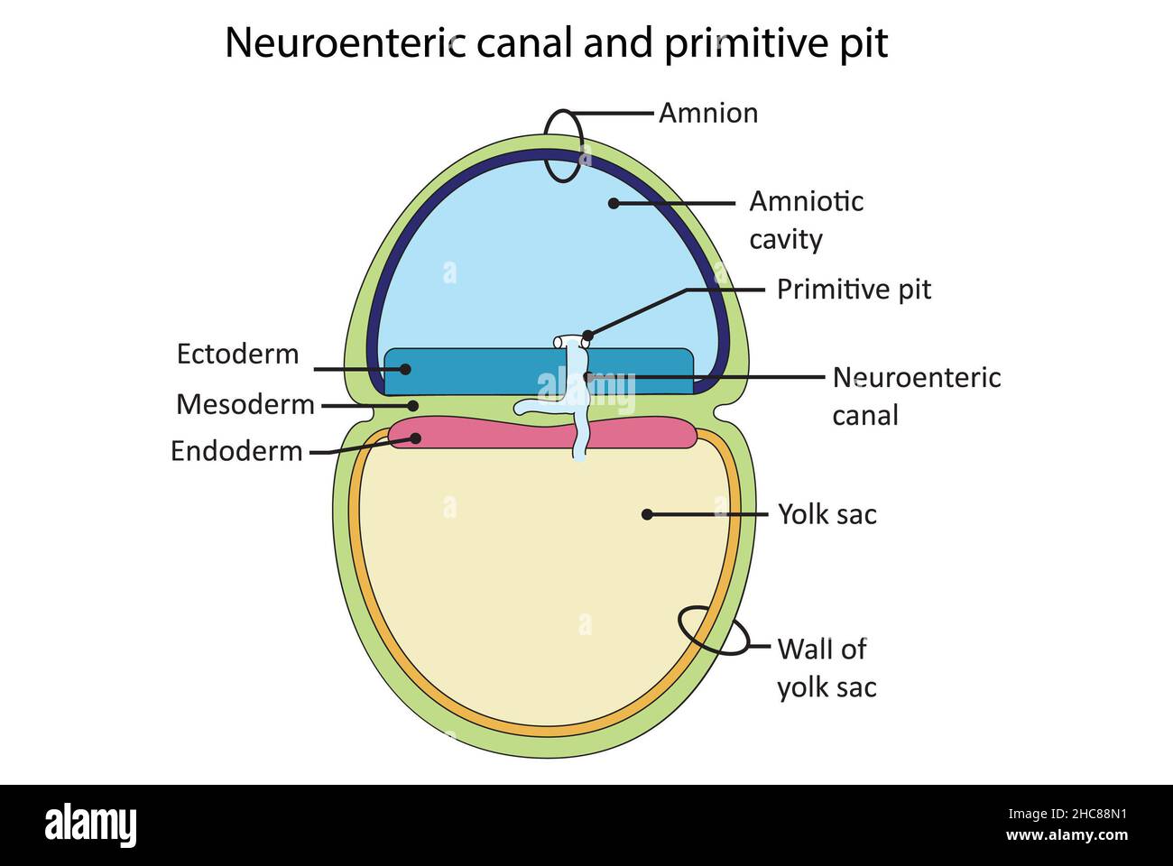
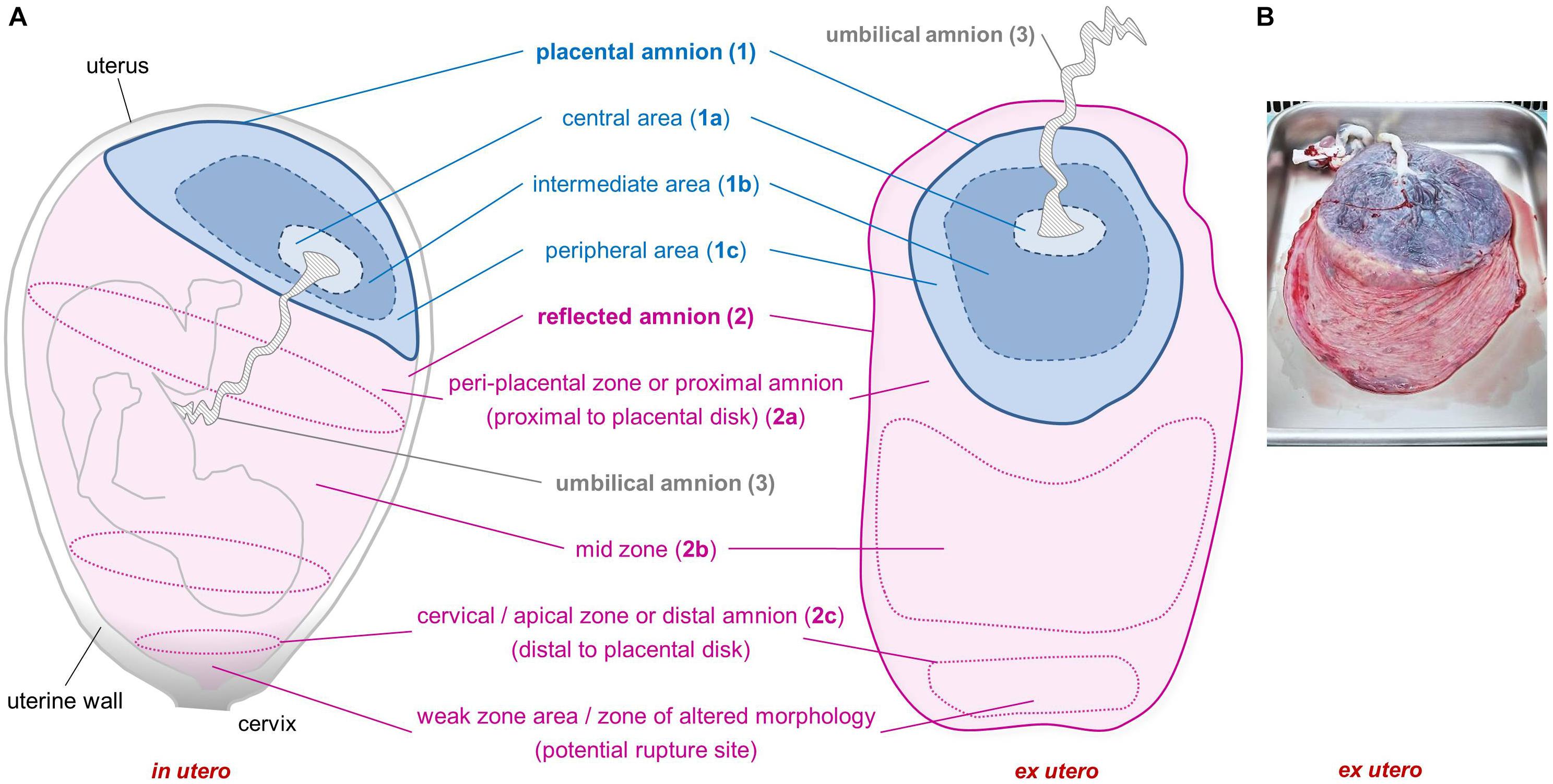
The placental amnion [ figure 1a (1)] covers the placenta, and the reflected amnion [ figure 1a (2)] lines the uterine wall, thereby forming the. Thus, placenta is partly maternal and partly. Download scientific diagram | anatomy of human placenta. The placenta is a connection between foetal membrane and the inner uterine wall. In placenta percreta, villi reach the uterine serosa and/or invade. The amnion, umbilical cord, and fetal vessels are visible. Here’s a detailed description of the umbilical cord, placenta, amniotic sac, and amniotic fluid, including their functions in the. The fetus developing within the amniotic sac is surrounded and encased by trophoblast cells in the placenta and chorion membrane (red). In placenta increta, villi penetrate deeper into the myometrium.

Uterus Placenta Amniotic Sac
In placenta increta, villi penetrate deeper into the myometrium. Here’s a detailed description of the umbilical cord, placenta, amniotic sac, and amniotic fluid, including their functions in the. Download scientific diagram | anatomy of human placenta. Thus, placenta is partly maternal and partly. The fetus developing within the amniotic sac is surrounded and encased by trophoblast cells in the placenta.

Uterus Placenta Amniotic Sac
Thus, placenta is partly maternal and partly. The fetus developing within the amniotic sac is surrounded and encased by trophoblast cells in the placenta and chorion membrane (red). The amnion, umbilical cord, and fetal vessels are visible. The placenta is a connection between foetal membrane and the inner uterine wall. Here’s a detailed description of the umbilical cord, placenta, amniotic.

Placenta Amniotic Sac Diagram
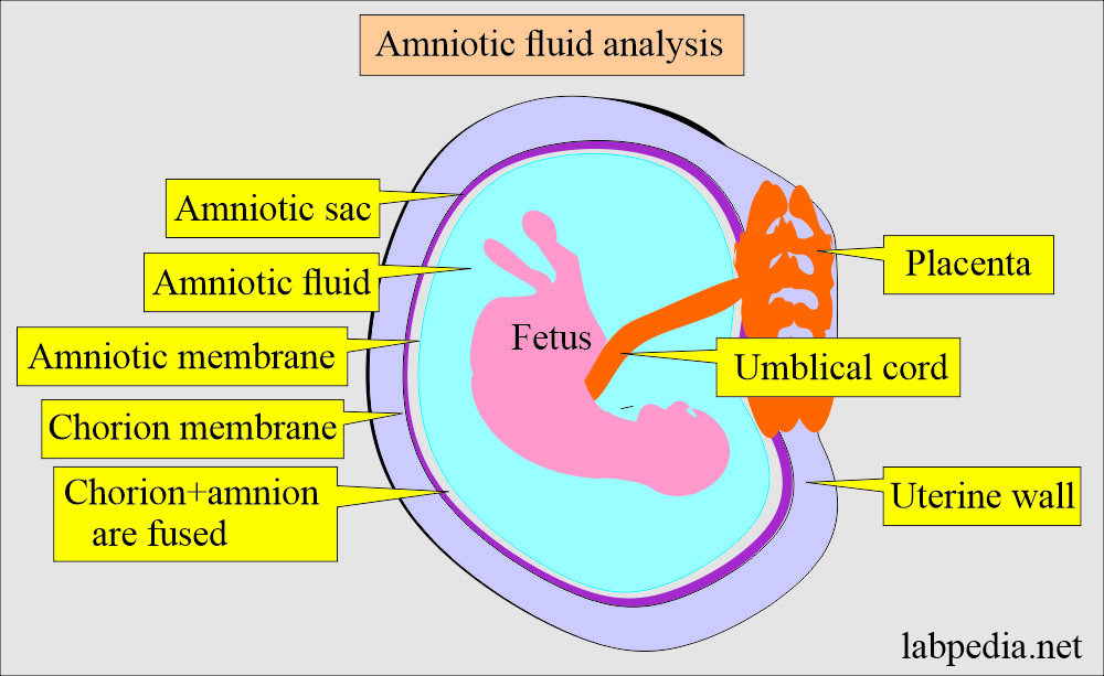
The fetus developing within the amniotic sac is surrounded and encased by trophoblast cells in the placenta and chorion membrane (red). The placenta is a connection between foetal membrane and the inner uterine wall. In placenta increta, villi penetrate deeper into the myometrium. Here’s a detailed description of the umbilical cord, placenta, amniotic sac, and amniotic fluid, including their functions.

Amniotic Sac Diagram
In placenta percreta, villi reach the uterine serosa and/or invade. Here’s a detailed description of the umbilical cord, placenta, amniotic sac, and amniotic fluid, including their functions in the. The placenta is a connection between foetal membrane and the inner uterine wall. In placenta increta, villi penetrate deeper into the myometrium. The fetus developing within the amniotic sac is surrounded.
Amniotic Sac Diagram
In placenta increta, villi penetrate deeper into the myometrium. Download scientific diagram | anatomy of human placenta. The placenta is a connection between foetal membrane and the inner uterine wall. The placental amnion [ figure 1a (1)] covers the placenta, and the reflected amnion [ figure 1a (2)] lines the uterine wall, thereby forming the. In placenta percreta, villi reach.
Placenta Amniotic Sac Diagram
The placental amnion [ figure 1a (1)] covers the placenta, and the reflected amnion [ figure 1a (2)] lines the uterine wall, thereby forming the. The fetus developing within the amniotic sac is surrounded and encased by trophoblast cells in the placenta and chorion membrane (red). Here’s a detailed description of the umbilical cord, placenta, amniotic sac, and amniotic fluid,.
Placenta And Amniotic Sac Diagram
Thus, placenta is partly maternal and partly. In placenta increta, villi penetrate deeper into the myometrium. The fetus developing within the amniotic sac is surrounded and encased by trophoblast cells in the placenta and chorion membrane (red). The placenta is a connection between foetal membrane and the inner uterine wall. The amnion, umbilical cord, and fetal vessels are visible.

Amniotic Sac Diagram
Download scientific diagram | anatomy of human placenta. The fetus developing within the amniotic sac is surrounded and encased by trophoblast cells in the placenta and chorion membrane (red). In placenta increta, villi penetrate deeper into the myometrium. The placental amnion [ figure 1a (1)] covers the placenta, and the reflected amnion [ figure 1a (2)] lines the uterine wall,.
Uterus Placenta Amniotic Sac
The fetus developing within the amniotic sac is surrounded and encased by trophoblast cells in the placenta and chorion membrane (red). Here’s a detailed description of the umbilical cord, placenta, amniotic sac, and amniotic fluid, including their functions in the. The amnion, umbilical cord, and fetal vessels are visible. In placenta increta, villi penetrate deeper into the myometrium. In placenta.

Amniotic Sac Diagram
Download scientific diagram | anatomy of human placenta. The placental amnion [ figure 1a (1)] covers the placenta, and the reflected amnion [ figure 1a (2)] lines the uterine wall, thereby forming the. The amnion, umbilical cord, and fetal vessels are visible. In placenta percreta, villi reach the uterine serosa and/or invade. In placenta increta, villi penetrate deeper into the.
The Fetus Developing Within The Amniotic Sac Is Surrounded And Encased By Trophoblast Cells In The Placenta And Chorion Membrane (Red).
The placental amnion [ figure 1a (1)] covers the placenta, and the reflected amnion [ figure 1a (2)] lines the uterine wall, thereby forming the. The placenta is a connection between foetal membrane and the inner uterine wall. Thus, placenta is partly maternal and partly. In placenta percreta, villi reach the uterine serosa and/or invade.
Here’s A Detailed Description Of The Umbilical Cord, Placenta, Amniotic Sac, And Amniotic Fluid, Including Their Functions In The.
Download scientific diagram | anatomy of human placenta. In placenta increta, villi penetrate deeper into the myometrium. The amnion, umbilical cord, and fetal vessels are visible.