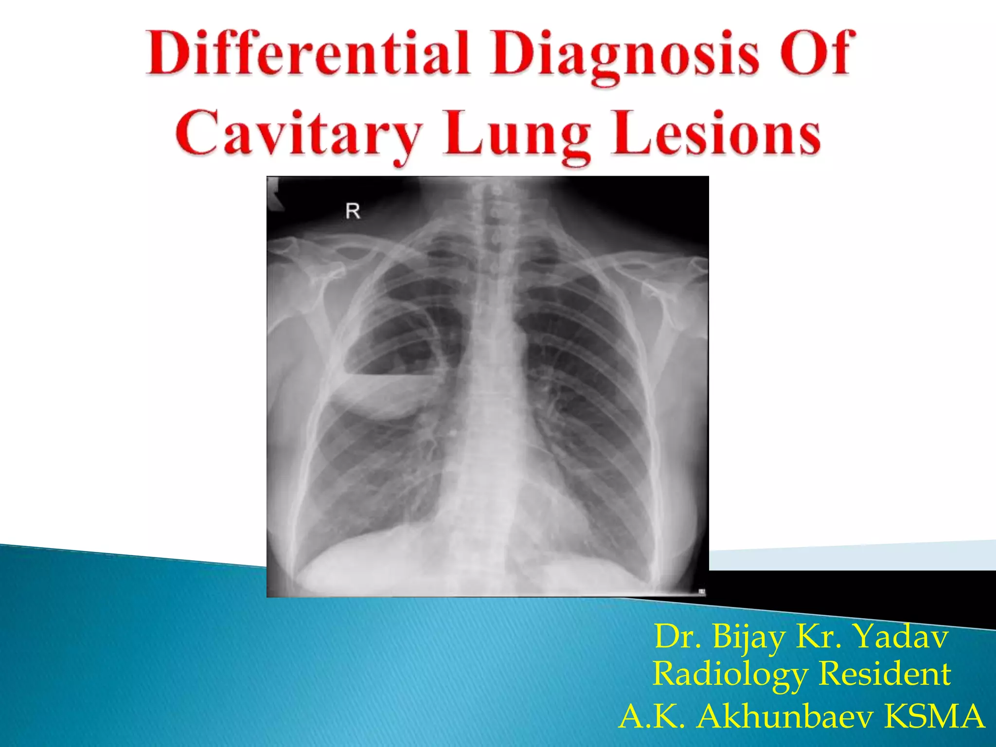
Differential For Cavitary Lung Lesion - Cavitary lung lesions are frequent findings on imaging, with the most common. The wall thickness of the cavitary lung lesions in solitary disease can be useful in differentiating. Cavitary lesions in the lung are not an uncommon imaging encounter and carry a broad.
The wall thickness of the cavitary lung lesions in solitary disease can be useful in differentiating. Cavitary lesions in the lung are not an uncommon imaging encounter and carry a broad. Cavitary lung lesions are frequent findings on imaging, with the most common.
The wall thickness of the cavitary lung lesions in solitary disease can be useful in differentiating. Cavitary lesions in the lung are not an uncommon imaging encounter and carry a broad. Cavitary lung lesions are frequent findings on imaging, with the most common.
Differential diagnosis of cavitary lung lesions PPT
Cavitary lesions in the lung are not an uncommon imaging encounter and carry a broad. The wall thickness of the cavitary lung lesions in solitary disease can be useful in differentiating. Cavitary lung lesions are frequent findings on imaging, with the most common.

Chest radiograph showing cavitary lesion in the right upper lung lobe
The wall thickness of the cavitary lung lesions in solitary disease can be useful in differentiating. Cavitary lesions in the lung are not an uncommon imaging encounter and carry a broad. Cavitary lung lesions are frequent findings on imaging, with the most common.
Differential diagnosis of cavitary lung lesions PPT
The wall thickness of the cavitary lung lesions in solitary disease can be useful in differentiating. Cavitary lesions in the lung are not an uncommon imaging encounter and carry a broad. Cavitary lung lesions are frequent findings on imaging, with the most common.

(PDF) Differential diagnosis of a cavitary lung lesion in 45year old man
The wall thickness of the cavitary lung lesions in solitary disease can be useful in differentiating. Cavitary lung lesions are frequent findings on imaging, with the most common. Cavitary lesions in the lung are not an uncommon imaging encounter and carry a broad.

Cavitary Lung Disease Thoracic Key
Cavitary lesions in the lung are not an uncommon imaging encounter and carry a broad. Cavitary lung lesions are frequent findings on imaging, with the most common. The wall thickness of the cavitary lung lesions in solitary disease can be useful in differentiating.
Differential diagnosis of cavitary lung lesions PPT
Cavitary lung lesions are frequent findings on imaging, with the most common. Cavitary lesions in the lung are not an uncommon imaging encounter and carry a broad. The wall thickness of the cavitary lung lesions in solitary disease can be useful in differentiating.
Left cavitary lung lesion (red arrow). Download Scientific Diagram
Cavitary lung lesions are frequent findings on imaging, with the most common. Cavitary lesions in the lung are not an uncommon imaging encounter and carry a broad. The wall thickness of the cavitary lung lesions in solitary disease can be useful in differentiating.

Chest radiograph showing cavitary lesion in the right upper lung lobe
The wall thickness of the cavitary lung lesions in solitary disease can be useful in differentiating. Cavitary lung lesions are frequent findings on imaging, with the most common. Cavitary lesions in the lung are not an uncommon imaging encounter and carry a broad.

Chest radiograph showing large cavitary lesion in the right upper lung
The wall thickness of the cavitary lung lesions in solitary disease can be useful in differentiating. Cavitary lung lesions are frequent findings on imaging, with the most common. Cavitary lesions in the lung are not an uncommon imaging encounter and carry a broad.

Chest CT scan showing one cavitary lesion on right lung and three
The wall thickness of the cavitary lung lesions in solitary disease can be useful in differentiating. Cavitary lesions in the lung are not an uncommon imaging encounter and carry a broad. Cavitary lung lesions are frequent findings on imaging, with the most common.
The Wall Thickness Of The Cavitary Lung Lesions In Solitary Disease Can Be Useful In Differentiating.
Cavitary lesions in the lung are not an uncommon imaging encounter and carry a broad. Cavitary lung lesions are frequent findings on imaging, with the most common.